| 目录: 28518 |
| 产品名称: Her3(DM175) Rabbit Monoclonal Antibody |
| 基因符号: HER3; ERBB3 |
| 描述: Her3 antibody(DM175) Rabbit Monoclonal Antibody |
| 背景: This gene encodes a member of the epidermal growth factor receptor (EGFR) family of receptor tyrosine kinases. This membrane-bound protein has a neuregulin binding domain but not an active kinase domain. It therefore can bind this ligand but not convey the signal into the cell through protein phosphorylation. However; it does form heterodimers with other EGF receptor family members which do have kinase activity. Heterodimerization leads to the activation of pathways which lead to cell proliferation or differentiation. Amplification of this gene and:or overexpression of its protein have been reported in numerous cancers; including prostate; bladder; and breast tumors. Alternate transcriptional splice variants encoding different isoforms have been characterized. One isoform lacks the intermembrane region and is secreted outside the cell. This form acts to modulate the activity of the membrane-bound form. Additional splice variants have also been reported; but they have not been thoroughly characterized. |
| 经过测试的应用: ELISA; Flow Cyt |
| 推荐稀释度: ELISA 1:5000-10000; Flow Cyt 1:100 |
| 种属反应性: Rabbit |
| 亚型: Rabbit IgG |
| 纯化: Purified from cell culture supernatant by affinity chromatography |
| 种属反应性: Human HER3 |
| 成分: Lyophilized from sterile PBS, pH 7.4. 5 % – 8% trehalose is added as protectants before lyophilization. |
| 储存和运输: Store at -20°C to -80°C for 12 months in lyophilized form. After reconstitution, if not intended for use within a month, aliquot and store at -80°C (Avoid repeated freezing and thawing). |

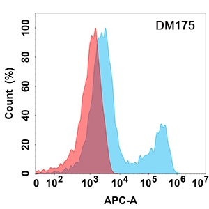
Figure 1. Flow cytometry analysis with Her3 (DM175) on Expi293 cells transfected with human Her3 (Blue histogram) or Expi293 transfected with irrelevant protein (Red histogram). |


