|
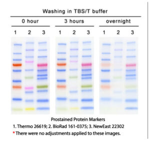
5. High Membrane Binding Affinity The membrane (PVDF or NC) after transferring during western blot experiment needs to undergo blocking, primary and secondary antibody incubation, and a multi-step washing process in between. It can take as short as 3 hours or even overnight. In a controlled experiment, our marker and ones of Thermo and Biorad are subjected to 3 hours to overnight washing with TBST washing buffer. After 3 hours, all bands of our products are very clear while the brightness of Thermo’s 10 kD band has been extremely weak and Biorad’s 25K band has completely disappeared, and its 10 KD band is also extremely weak. After overnight washing, only 25K and 20K of our marker are weakened, and the rest of the bands are still very clear. In comparison, Thermo’s 10 KD band is almost completely disappeared, and the two red bands is very faint. More than half of the bands for Biorad product disappears or becomes very faint. The membrane (PVDF or NC) after transferring during western blot experiment needs to undergo blocking, primary and secondary antibody incubation, and a multi-step washing process in between. It can take as short as 3 hours or even overnight. In a controlled experiment, our marker and ones of Thermo and Biorad are subjected to 3 hours to overnight washing with TBST washing buffer. After 3 hours, all bands of our products are very clear while the brightness of Thermo’s 10 kD band has been extremely weak and Biorad’s 25K band has completely disappeared, and its 10 KD band is also extremely weak. After overnight washing, only 25K and 20K of our marker are weakened, and the rest of the bands are still very clear. In comparison, Thermo’s 10 KD band is almost completely disappeared, and the two red bands is very faint. More than half of the bands for Biorad product disappears or becomes very faint. | |
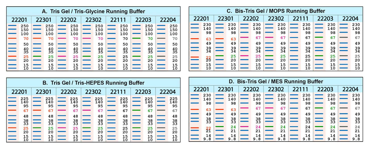
6. Migration Patterns The migration of the prestained marker proteins during electrophoresis depends not only on the proteins themselves but also on the coupled dyes. Therefore, the markers usually show different migration patterns in different electrophoresis systems. The MWs of them are originally calibrated in Tris gel/Tris-Glycine buffer system (Table A). The shifts of the markers in other electrophoresis systems are shown in Table B-D for references. We highly recommend to specifically calibrate their MWs by unstained protein MW standard in corresponding systems other than Tris gel/Tris-Glycine buffer. The migration of the prestained marker proteins during electrophoresis depends not only on the proteins themselves but also on the coupled dyes. Therefore, the markers usually show different migration patterns in different electrophoresis systems. The MWs of them are originally calibrated in Tris gel/Tris-Glycine buffer system (Table A). The shifts of the markers in other electrophoresis systems are shown in Table B-D for references. We highly recommend to specifically calibrate their MWs by unstained protein MW standard in corresponding systems other than Tris gel/Tris-Glycine buffer. | |
| Usage: The protein ladder comes ready to use in gel loading buffer. After thawing and mixing, load approximately 3-5 µL of protein marker per lane. Do NOT heat, dilute, or add reducing agents before loading |
| Detection Method: Colorimetric |
| Molecular Weight Range: 10-250 KD |
| Stain Type : Red and Blue |
| Constituents : 62.5 mM Tris‐HCl (pH 7.5 at 25°C), 1 mM EDTA, 2% (w/v) SDS, 10 mM DTT and 30% (v/v) glycerol |
| Storage : 4 °C for up to 1 year while -20°C and -80°C for up to 3 and 10 years, respectively. Avoid repeated freezing and thawing |


 NewEast Biosciences Prestained Protein Marker is as accurate as the current non-prestained ladders on the market (Compare lanes 1 and 2 with ours lane 6), but has superior MW precision in comparison with the major market suppliers, ie. Biorad and Thermo (Compare lanes 3, 4 and 5 with ours lane 6). Biorad ladder has two inaccurate bands of 37K and 75K while Thermo prestained products have inconsistent 35K and 25K. Even more, Thermo has many inconsistencies in the molecular weight between its prestained and non-prestained protein markers.
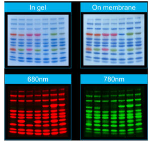
 The images are produced from a single controlled experiment. All bands except orange ones are well compatible with near-infrared (NIR) fluorescent system. Note that the red band in Thermo’s product cannot be scanned by NIR fluorescence either. The 货号: of protein markers (left to right) is 22201, 22301, 22202, 22302, 22101, 22203 and 22204.
 The membrane (PVDF or NC) after transferring during western blot experiment needs to undergo blocking, primary and secondary antibody incubation, and a multi-step washing process in between. It can take as short as 3 hours or even overnight. In a controlled experiment, our marker and ones of Thermo and Biorad are subjected to 3 hours to overnight washing with TBST washing buffer. After 3 hours, all bands of our products are very clear while the brightness of Thermo’s 10 kD band has been extremely weak and Biorad’s 25K band has completely disappeared, and its 10 KD band is also extremely weak. After overnight washing, only 25K and 20K of our marker are weakened, and the rest of the bands are still very clear. In comparison, Thermo’s 10 KD band is almost completely disappeared, and the two red bands is very faint. More than half of the bands for Biorad product disappears or becomes very faint.
 The migration of the prestained marker proteins during electrophoresis depends not only on the proteins themselves but also on the coupled dyes. Therefore, the markers usually show different migration patterns in different electrophoresis systems. The MWs of them are originally calibrated in Tris gel/Tris-Glycine buffer system (Table A). The shifts of the markers in other electrophoresis systems are shown in Table B-D for references. We highly recommend to specifically calibrate their MWs by unstained protein MW standard in corresponding systems other than Tris gel/Tris-Glycine buffer.